2018-2019 BMES-Medtronic Student Design Competition Winners
2019'S TOPIC OF FOCUS WAS DIGITAL IMAGING.
First Place was awarded $1,500 with $1,500 in travel reimbursement
Second Place was awarded $1,000 with $750 in travel reimbursement
Third Place was awarded $500 with $500 in travel reimbursement
 FIRST PLACE WENT TO THE TEAM REPRESENTING THE UNIVERSITY OF FLORIDA WITH ITS INNOVATIVE DESIGN:
A positive diagnosis of prostate cancer is currently obtained only via pathology examination of biopsied samples whereby samples are observed to contain cancerous cells. However, there is a 21-47% rate of false-negative biopsies. These false-negative biopsy reports unnecessarily delay initiation of treatment which, in turn, may give time for localized prostate cancer to metastasize and spread to other organs and organ systems, reducing options, complicating treatment and worsening outcomes.
Our team designed an ultrasound-guided prostate biopsy mixed reality simulation, in collaboration with UF Center for Safety, Simulation & Advanced Learning Technologies (CSSALT), with the aim of developing a cost-effective and portable educational simulator that would allow clinicians to assess their performance and improve their prostate biopsy techniques. This device combined both virtual components, such as ultrasound images and anatomical diagram guides, with tracked physical components, such as a physical prostate, needle gun and ultrasound probe. The simulator used an existing commercial tracking system in conjunction with 3D visualization to provide clinicians a means to practice and refine their technique in a low-risk, low-stress environment. More importantly, the simulator provided real-time feedback of biopsy locations and probe positioning in relation to both physically and virtually simulated prostate. The simulator software was based on the SMARTS-SDK (developed by CSSALT) and implemented in the Unity3D game engine.
As of now, our team has passed our design to UF CSSALT. UF CSSALT has already adopted our design to be used in international studies, which have shown initial positive results in reducing false negative rates and improving accuracy of biopsies.

SECOND PLACE WAS AWARDED TO THE TEAM REPRESENTING ARIZONA STATE UNIVERSITY WITH THEIR DESIGN:
Skin cancer is currently the most prevalent type of cancer in the United States: about 9,500 individuals are diagnosed daily, however, skin cancer is also one of the most controllable and preventable types of cancer. Our solution is a mobile app with the following features: User-friendly imaging technology, an active community network of medical professionals and patients, a crowd-sourced image database for different skin cancers, and an easily accessible educational component to raise awareness and provide patients more resources on skin cancer detection and prevention.
“Know Your Skin” uniquely achieve several important goals: First, it uses a more comprehensive approach to evaluate several common skin cancers, rather than just focusing on melanoma or one single type of cancer. Second, it includes added features of a collective database and community to improve the quality of self-examination. Users will be able to cross-reference their potentially malignant lesions with other patients to gain greater insight into their risk profile. Finally, “Know Your Skin” allows for greater personalization and customizability through the combination of all the features this app provides.

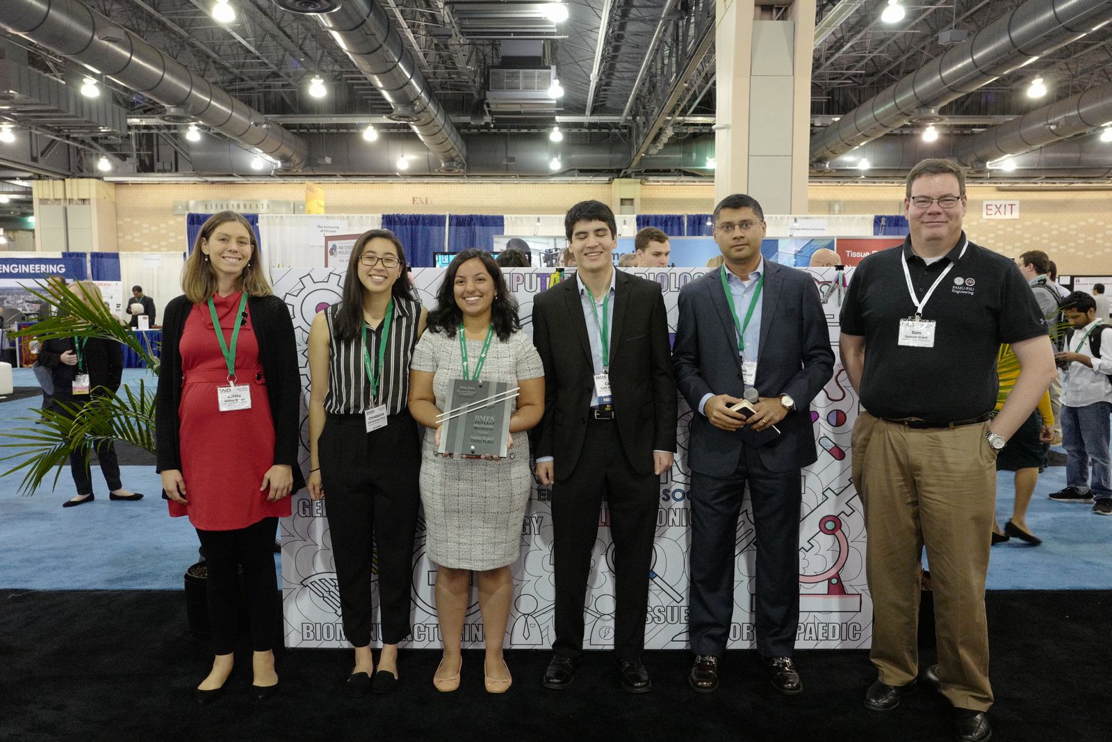
THIRD PLACE WAS AWARDED TO THE TEAM REPRESENTING GEORGE WASHINGTON UNIVERSITY WITH THEIR DESIGN:
Our blur-reducing medical imaging technology is a sensor-software coupled system that informs medical technicians of the optimal time to capture a medical image (such as an X-Ray) in order to minimize image blur/distortion and thereby reduce unnecessary image retakes and ionizing radiation exposure.
Our anticipated regulatory pathway will be an FDA 510k, which will introduce our device to market in a satisfactory manner. Due to our device’s lack of safety concerns, there will be no need to undergo clinical trials associated with more potentially dangerous innovations. Our device will only act as a safety buffer between image capture and triggering event to take an X-ray.
We will be selling our end product at $220 per unit, and the production cost is approximately $120 per unit. On the sale of each product, there will be a profit of approximately $100. At 2700 products sold, we break even with our startup costs. We plan to market the product as a two-piece bundle of the software code and hardware sensor to physicians and technicians who are purchasing this as an add-on to their hospital or clinic’s existing X-ray machine. Our end goal is acquisition by a larger medical device company that could fully integrate our technology into their X-ray machines.